
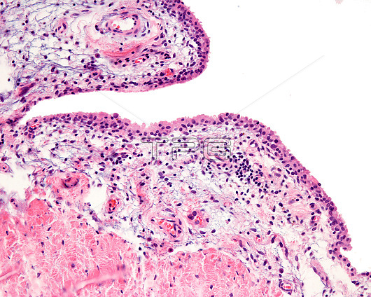
Light micrograph of a human synovial membrane (innermost part of the joint capsule). It delimits the joint cavity of the diarthrosis. A band of synovial cells is concentrated in the vicinity of the lumen of the joint cavity, adopting an appearance reminiscent of a lining epithelium; however, these synovial cells are connective tissue cells that form a discontinuous lining. Deeper is a large space occupied by loose connective tissue with abundant blood vessels containing red cells in the lumen. In the lower frame, a reddish band is observed, rich in collagen fibres that corresponds to the fibrous capsule.
| px | px | dpi | = | cm | x | cm | = | MB |
Details
Creative#:
TOP26624879
Source:
達志影像
Authorization Type:
RM
Release Information:
須由TPG 完整授權
Model Release:
N/A
Property Release:
N/A
Right to Privacy:
No
Same folder images:
 Loading