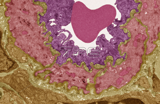
High endothelial venule. Coloured transmission electron micrograph (TEM) of the tall cuboidal endothelial cells (upper layer) lining a high endothelial venule (HEV). Venules are little veins that transport deoxygenated blood from the capillary beds to the veins. Endothelial cells line the entire circulatory system but HEVs are found specifically in lymph nodes, tonsils and Peyer's patches (lymphoid tissue in the small intestine). The special cuboidal endothelial cells, along with receptors, allow leukocytes (white blood cells) to gain entry into the lymph node via the blood. Smooth muscle cells (red) and connective tissue surround the endothelium. A single red blood cell is present in the lumen of the venule. Magnification: x3000 when printed 10 centimetres wide.
| px | px | dpi | = | cm | x | cm | = | MB |
Details
Creative#:
TOP25847318
Source:
達志影像
Authorization Type:
RM
Release Information:
須由TPG 完整授權
Model Release:
N/A
Property Release:
N/A
Right to Privacy:
No
Same folder images:

 Loading