
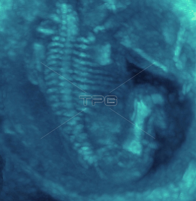
Foetus skeleton. Coloured 3-D ultrasound scan of part of the skeleton of a foetus. The spine is seen from upper left to lower centre, with ribs on either side. The pale blue shape at lower right is the femur (thigh bone) of the upper leg. Ultrasound scanning is a diagnostic technique that sends high-frequency sound waves into the body via a transducer. The returning echoes are recorded and used to build an image of an internal structure. Foetal ultrasound scanning is routine during pregnancy. A technique called Maximum and Minimum Intensity Projection (MIP) is used, in association with 3-D ultrasound, to visualize the bones of a developing foetus.
| px | px | dpi | = | cm | x | cm | = | MB |
Details
Creative#:
TOP10222401
Source:
達志影像
Authorization Type:
RM
Release Information:
須由TPG 完整授權
Model Release:
N/A
Property Release:
N/A
Right to Privacy:
No
Same folder images:
 Loading