
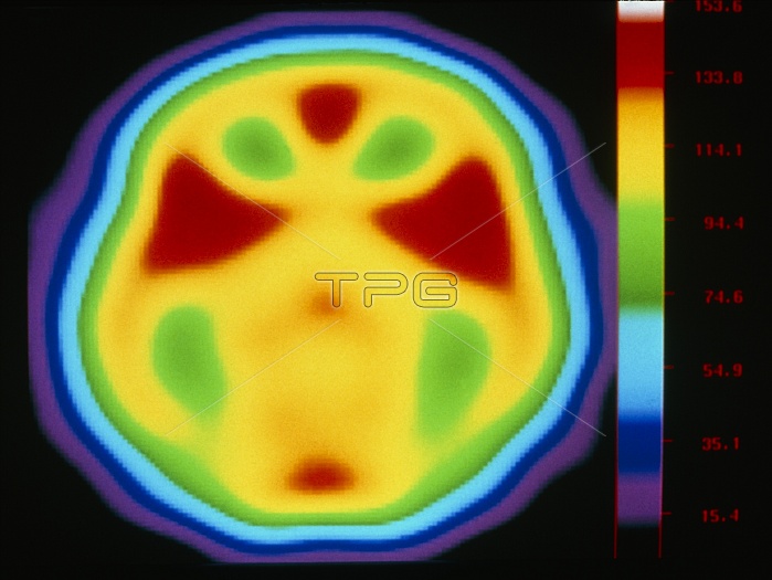
Brain's auditory centre. Coloured positron emission tomography (PET) scan of the brain during a listening exercise. The exercise involved unfocused listening to background noise. In this axial (horizontal) scan the front of the brain is at top. The scan shows oxygen and water levels from low (blue) to high (red). These levels correspond to brain activity, the highest levels being in the auditory cortex (red triangles) of the two temporal lobes. PET scans use radioactively-labelled substances introduced into the blood to view metabolic activity. See P335/035 for a PET scan taken during focused listening.
| px | px | dpi | = | cm | x | cm | = | MB |
Details
Creative#:
TOP10219330
Source:
達志影像
Authorization Type:
RM
Release Information:
須由TPG 完整授權
Model Release:
N/A
Property Release:
N/A
Right to Privacy:
No
Same folder images:
 Loading