
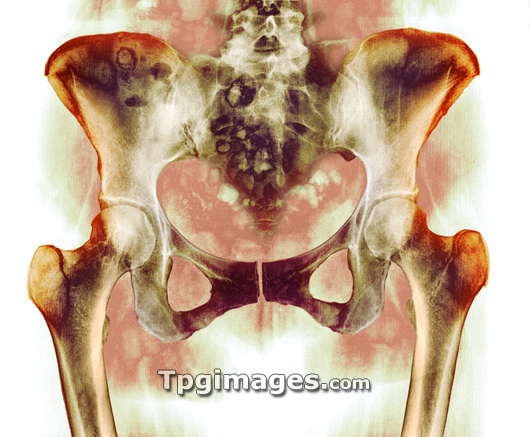
Healthy hip bones. Coloured X-ray of the pelvis of a 49 year old woman, showing bones of the lower spine, hips and thighs. The two hip joints are at centre right and left in this frontal view; these ball-and-socket joints provide the mobility needed for walking and running. The socket is a cavity in this part of the pelvis. The ball of the hip joint is the head of the femur (thigh bone); this head is seen joined by a neck to the rest of the femur, the long bone descending down lower left and right sides. The base of the spine is at top centre, and the distinctive loops at the base of the pelvis are also seen. The female pelvis is wider and shallower than the male pelvis in order to create a wide birth canal.
| px | px | dpi | = | cm | x | cm | = | MB |
Details
Creative#:
TOP03220809
Source:
達志影像
Authorization Type:
RM
Release Information:
須由TPG 完整授權
Model Release:
N/A
Property Release:
N/A
Right to Privacy:
No
Same folder images:
 Loading