
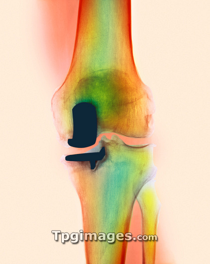
Partial knee joint replacement. Coloured X-ray of the knee of a 61 year old man (front view), showing a partial prosthesis (artificial joint, dark colour) replacing a damaged part of the knee joint. Osteoarthritis in this patient caused part of the old joint to lose its cartilage; healthy cartilage forms a hard surface between the bones reducing friction, and it is progressively lost due to this disease. The implant made of metal alloy attaches to the bottom of the femur (thigh bone, upper frame) and to the top of the tibia (shin bone, lower frame). It forms a flexible joint that can hinge like the old joint, relieving joint pain and immobility.
| px | px | dpi | = | cm | x | cm | = | MB |
Details
Creative#:
TOP03217836
Source:
達志影像
Authorization Type:
RM
Release Information:
須由TPG 完整授權
Model Release:
N/A
Property Release:
N/A
Right to Privacy:
No
Same folder images:
 Loading