
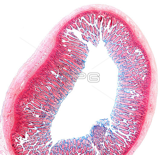
Light micrograph of a cross-section of the small intestine stained with the Muller-Mowry colloidal iron technique. Goblet cells appear as blue points in the epithelium lining the villi and the crypts of Lieberkuhn.
| px | px | dpi | = | cm | x | cm | = | MB |
Details
Creative#:
TOP29223588
Source:
達志影像
Authorization Type:
RM
Release Information:
須由TPG 完整授權
Model Release:
Not Available
Property Release:
Not Available
Right to Privacy:
No
Same folder images:
 Loading